Treating Common Foot Pain & Foot Deformities
Get Relief from Foot Pain at any of our 5 Tulsa Locations
Tulsa Foot Pain and Deformities Specialists
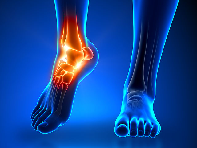
Foot pain may be caused by injuries, improper footwear, deformities, and biomechanical conditions that cause inflammation or stresses involving bones, ligaments or tendons in the foot. Infectious diseases, bacteria, fungi and viruses are also known to be a cause of foot pain. Delayed treatment of foot pain can lead to complications, chronic pain, disability and even arthritis of the affected foot. When pain begins to interfere with daily living, it is time to seek medical attention.

We understand that foot pain can be debilitating and cause unnecessary problems.
GET RELIEF TODAY
Get Relief from Common Foot Pain & Foot Deformities
7 reasons to see a podiatrist
Your feet are complex structures with many bones, ligaments, and tendons that work together to support your body and [read more]
5 ways to identify neuropathy in feet and ankles
Neuropathy, specifically of the feet and ankles, affects millions of people worldwide, often leading to pain, numbness, and a [read more]
Tori Record
Q: Why do you love working at Metro Tulsa Foot & Ankle Specialists? A: Since I can remember, [read more]
Subscribe to stay up-to-date on news and tips from us.
Bunion
Bunions are a common foot deformity caused by the big tow leaning toward the second toe, causing the bones to be thrown out of alignment, thus producing the “bunion’s bump.” Many people may unnecessarily suffer from the pain of the bunions for years before seeking treatment. Since bunion’s are a progressive disorder, over time the discomfort and characteristics of the bunion bump will become more prominent. Symptoms of discomfort are often aggravated when an individual is wearing shoes that crows their toes or spending long periods of time on your feet. Symptoms include:
- Pain and Soreness on and around the bunion bump
- Inflammation and Redness
- A burning sensation
- Possible Numbness
How are bunions treated?
Although bunions are often identified from prominent visibility, we may take x-rays to determine the degree of the deformity to ensure the best possible treatment plan. There are many non-surgical treatment options that we consider before planning surgery. These include making sure you are wearing the right kind of shoes, activity modifications to ensure you are giving your feet the rest they need, prescribing anti-inflammatory medication and icing the bunion to help reduce inflammation and pain, as well as custom orthotic devices to give your feet the support they need. After all non-surgical conditions are considered, if there has not been progress in the condition, we have many surgical specialists that will determine if surgery is the right treatment option for you. From there we will work with you to explain the surgical technique and recovery process so we can help reduce the pain and deformity the bunion has caused.
Bunionette
What is a bunionette?
A bunionette, also called Tailor’s bunion, describes the prominence of the fifth metatarsal bone at the base of the little toe. A bunionette is caused by an inherited faulty mechanical structure of the foot. This rare structure causes the fifth metatarsal bone to protrude outward, while the toe moves inward, creating a bump on the outside of the foot that becomes irritated when a shoe presses against it.
Symptoms include:
- Redness
- Swelling
- Pain
How is a bunionette treated?
Treatment for a bunionette begins with nonsurgical therapies including shoe modifications, padding, oral medications, injection therapy, and orthotic devices. Our surgical specialists may consider surgery if your pain continues despite previously used nonsurgical therapy approaches.
Cavus Foot (High-Arched Foot)
What is Cavus Foot?
Cavus foot describes a condition in which the foot has an extremely high arch causing an excessive amount of pressure to be placed on the ball and heel of the foot when walking or standing. Cavus foot is something that can develop at any age and is often caused by a neurologic disorder such as cerebral palsy, Charcot-Marie-Tooth disease, spina bifida, polio, muscular dystrophy or stroke. If Cavus foot is caused by a neurological disorder, is it like to get worse with time. Symptoms of Cavus Foot include:
- Hammertoes or claw toes
- Calluses on the ball, side or heel of the foot
- Pain when standing or walking
- An unstable foot due to heel tilting inward which can lead to ankle sprains
- Foot drop, or weakness of the muscles of the foot that lead to the foot-dragging when a step is taken
How is Cavus Foot treated?
Nonsurgical options to treat Cavus Foot are custom orthotic devices, shoe modifications, and bracing. In the event that nonsurgical treatment does not help to relieve your pain and improve the stability of your foot, surgery may be deemed necessary. In cases where an underlying neurological problem exists, surgery may be needed again in the future due to the progression of this disorder.
Ganglion Cyst
What is a ganglion cyst?
A ganglion cyst is a sac filled with fluid that originates from a tendon sheath or joint capsule. The term “ganglion” means knot and is used to describe the knot-like mass or lump that forms below the surface of the skin. Symptoms of a ganglion cyst are:
- A noticeable lump
- Tingling or burning, if the cyst is touching a nerve
- Dull pain or ache
- Difficulty wearing shoes due to irritation between the lump and the shoe.
How is a ganglion cyst treated?
One of our skilled surgeons will perform a thorough examination of your foot. The lump will be pressed in different ways and should move freely under the skin. Sometimes our team will remove a small amount of fluid from the cyst for evaluation. Treatment varies based on your pain in association with the cyst. If there is no pain and the cyst does not interfere with walking, our team may choose to monitor your cyst closely and suggest shoe modifications that may help provide you with comfort. At times, our team may choose to drain the fluid in house and then inject a steroid medication into the mass. If these treatment options are not appropriate for your cyst, the cyst may be surgically removed
Neuroma
What is a neuroma?
A neuroma is a thickening of nerve tissue that develops in various parts of the body, the most common being on the foot between the metatarsal bones. The thickening of the nerve is a result of compression and irritation of the nerve, and can eventually lead to permanent nerve damage.
Symptoms of neuroma are:
- Tingling, burning or numbness
- Pain
- A feeling that something is inside the ball of the foot
- A feeling that there is something in the shoe or a sock is bunched up
How is neuroma treated?
The best time to see our surgical specialists is early in the development of symptoms as this will lessen the need for invasive treatments. Nonsurgical options include padding, icing, orthotic devices, activity modifications, shoe modifications, medications, and injection therapy. Surgery may be considered for patients who have not responded adequately to nonsurgical treatments. Regardless of whether you have undergone surgical or nonsurgical treatment, our team will recommend long-term measures to help keep your symptoms from returning.
Arthritis
What is foot arthritis?
Foot arthritis can be defined as joint inflammation, and it is known to produce swelling and pain that could lead to deformity, loss of function and decreased ability to walk. Symptoms of foot arthritis include:
- Pain and stiffness in the joint
- Swelling in or near the joint
- Difficulty walking or bending the joint
How is foot arthritis treated?
Nonsurgical treatment for foot arthritis is oral medications, orthotic devices, bracing, immobilization, steroid injections and physical therapy. With advanced cases of foot arthritis, surgery may be the only option. The goal of surgery in this case would be to decrease your pain and improve function.
Calcaneal Apophysitis (Sever’s Disease)
What is Calcaneal Apophysitis (Sever’s Disease)?
Calcaneal Apophysitis is type of bone injury in which the heel’s growth plate becomes painful and inflamed. This is the most common cause of heel pain found in children between the ages of 8 and 14 due to the heel bone not being fully developed.
Symptoms of Calcaneal Apophysitis are:
- Pain in the back or bottom of the heel
- Limping
- Walking on toes
- Difficulty running, jumping or participating in usual activities or sports
- Pain when the sides of the heel are squeezed
- Tiredness
How is Calcaneal Apophysitis treated?
To better diagnose your child’s heel pain, we will start their exam by talking with you about your child’s medical history and recent activities. Our surgical specialist will then complete a physical exam and x-rays so that they are able to rule out more serious conditions. There are many nonsurgical approaches to treating heel pain such as reducing activity, supporting the heel, nonsteroidal anti-inflammatory medications, physical therapy and immobilization. Please be aware that this is a condition that may continue to plague your child even after it has been treated as the heel bone is still actively growing. If your child continues to have an excessive amount of discomfort, our staff would love to bring them back in for further review.
Haglund's Deformities
What is Hauglund’s deformity?
Huglund’s deformity refers to a bony enlargement found on the back of the heel. The issue for one with this deformity arises when the soft tissue by the Achilles tendon becomes irritated as it rubs against shoes, leading to painful bursitis. Symptoms of Haglund’s deformity can occur in one or both of the feet and include:
- A noticeable bump on the back of the heel
- Pain in the area where the Achilles tendon attaches to the heel
- Swelling in the back of the heel
- Redness near the inflamed tissue
How is Hauglund’s deformity treated?
Nonsurgical treatment is goaled at reducing the inflammation of the bursa, and includes nonsteroidal anti-inflammatory drugs, exercises, heel lifts, heel pads, shoe modifications, physical therapy, immobilization, and orthotic devise. When nonsurgical treatment does not provide adequate pain relief, surgery may be necessary.
Gout
What is gout?
Gout refers to a buildup of uric acid in the tissues or a joint, though it is most commonly found in the joint of the big toe. Uric acid is present in the blood and eliminated through urine, but with individuals who have gout the uric acid accumulates and crystallizes in the joints. Gout is sensitive to temperature changes, as cooler temperatures cause uric acid to turn into crystals. Since the toe is further from the heart, it is a cooler portion of the body and is thus more likely to be a target of gout. Symptoms of gout include:
- Intense pain that comes on suddenly, often in the middle of the night or upon arising
- Signs of inflammation, such as redness, swelling and warmth over the joint.
How is gout treated?
Initial treatment of a gout attack includes medications or injections that can be used to treat pain, dietary restrictions, fluids, and immobilization of the foot. Typically, a gout attack resolves in three to ten days with treatment. If gout symptoms continue to plague you, daily medication may help limit repeated episodes.
Foot Drop
What is foot drop?
Foot drop refers to individuals who are unable to lift the front of their foot off the ground when walking. Due to this, these individuals often scuff or drag their foot. This condition is generally caused by nerve or muscle disorders, or by an underlying central nervous system disorder.
How is foot drop treated?
Treatment for foot drop may include braces, physical therapy and electrical nerve stimulation, though in some cases surgery might be required.
Flexible Flatfoot
What is flexible flatfoot?
Flexible flatfoot is one of the most common forms of flatfoot. Flexible flatfoot usually begins in childhood and continues into adulthood. Most often this deformity occurs in both feet and gets worse with age and can cause the tendons and ligaments of the arch to stretch or tear. The term “flexible” comes from the fact that when you are standing, the foot is flat. Then upon sitting the arch returns. Symptoms of flexible flatfoot include:
- Pain in the heel, arch, ankle or along the outside of the foot
- Rolled-in ankle (overpronation)
- Pain along the shin bone (shin splint)
- General aching or fatigue in the foot or leg
- Low back, hip or knee pain.
How is flexible flatfoot treated?
Nonsurgical treatment options for flexible flatfoot include activity modifications, weight loss, orthotic devices, immobilization, nonsteroidal anti-inflammatory drugs, physical therapy, shoe modifications and ankle foot orthoses devices. In rare cases where pain is unable to be controlled by other treatments, surgery may be necessary. There are many surgical techniques that can help correct this deformity. Our specialists will work with you to find the best path for treatment that will not only relieve your symptoms but improve your foot function as well.
Turf Toe
What is turf toe?
Turf toe is a sprain to the big toe resulting from an injury during sports activities, or repeated jamming of ones big toe over time. The name “turf toe” is derived from the fact that this injury is common among athletes who play on artificial turf.
Signs and symptoms of turf toe include:
- Pain
- Swelling
- Limited joint movement
How is turf toe treated?
Turf toe nonsurgical treatment options include rest, ice, compression and elevation (also known as RICE), as well as changing to a more structured footwear.
Soft Tissue Biopsy
What is a soft-tissue biopsy?
A soft-tissue biopsy is removal of a small sample of soft tissue that can be placed under microscope for examination. Soft-tissue is the skin, fat, muscle and tendons that surround, connect and support other tissues. Acquiring this sample of tissue takes only a few minutes and will help our team reach an accurate diagnosis for your condition. At MTFAS we offer three different types of biopsies. All are performed after we numb the area. These types are:
- Shave biopsy – A thin piece of tissue is shaved off.
- Punch biopsy – A tiny round instrument is used to remove a miniscule amount out of the core of tissue. At times, bunch biopsies may require stitches.
- Incisional or excisional biopsy – a piece or an entire lesion is removed. Stitches are generally required for this type of biopsy.
Once the same is obtained, our staff will send the sample to be tested by a pathologist who specializes in soft tissue biopsy. If the area of concern was stitched together, we will make an appointment for you to return after several days for stitch removal. At this time we will go over the results of the biopsy with you.
Plantar Warts
What is a plantar wart?
A plantar wart is a small growth on the skin that develops when the skin is infected by a virus. These warts can be found anywhere on the foot, but they typically appear on the bottom (plantar side). A plantar wart can be a single – solitary wart, or it can be a mosaic wart which is defined as a cluster of warts. Symptoms of plantar warts include:
- Thickened skin, as a plantar wart usually resembles a callus
- Pain when walking or standing
- Tiny black dots often appear on the surface
How are plantar warts treated?
The goal of treating a plantar wart is to completely remove it. Our specialists may use topical or oral treatments, laser therapy, cryotherapy, acid treatments or surgery. Although many home remedies for warts exist, you should never try to remove warts yourself. This has been known to do more harm than good.


